A mechanical disorder ….. is …
characterized by …..
caused by ..etiology or predisposing factors
resulting in a pathological feature (structural change or functional change) or clinical feauture
Sometimes complicated by ….
Diagnosis is suspected clinically by … and confirmed by ….
Imaging includes the use of
Treatment options depend on …. but includes …..
Etymology if available
Principles
Pump Failure
Heart Failure Kerley B lines |
| 42545c01.800 heart cardiac lungs secondary lobules interstitium interlobular septa thickening fx Kerley B lines dx CHF congestive heart failure plain film CXR Davidoff MD |
Narrowing Stenosis
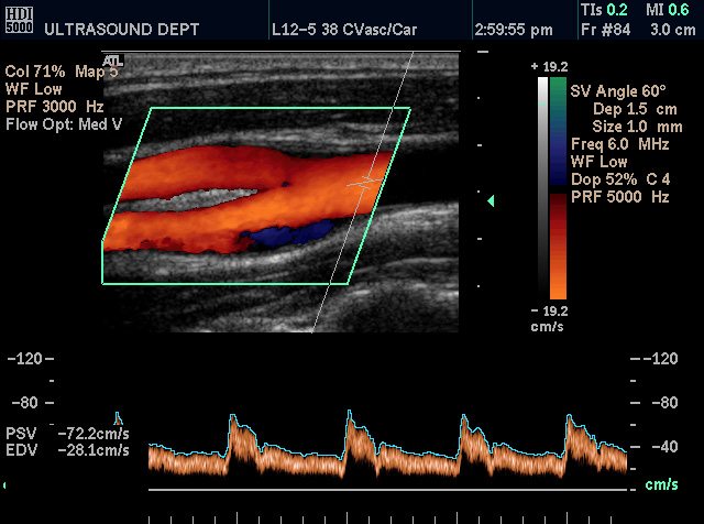
Carotid Artery – small region of stenosis |
| This color flow doppler US of the neck shows a carotid artery with bifurcation into internal and external carotid vessels. The small area of turbulence is noted by a blue area at the bifurcation.
Courtesy Philips Medical Systems 33288 |

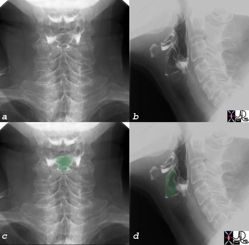
Normal and Tracheal Stenosis |
| 49838d01 50 year old female with respiratory difficulty trachea bronchi rectus abdominis muscle compression fractures kyphosis dwarf dwarfism right aortic arch tracheomalacia tracheal stenosis rectus abdominis muscle hypertrophy Davidoff MD Courtesy Ashley Davidoff MD CTscan 49838 49838c01 49838c02 49838c03 49838c04 49838c05 shape size position character growth |
Esophagus Extrinsic Pressure
Aberrant Right Subclavian artery Causing Compression of Esophagus |
| 16376c01 aorta aberrant origin of right subclavian artery as the last vessel off the left aortic arch congenital growth position esophagus CTscan Davidoff MD |
Obstruction
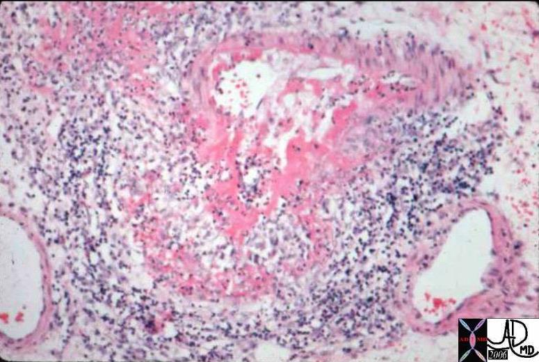
Polyarteritis Nodosa |
| Occlusive Vascular Disease: Causes of Vascular Occlusion – Polyarteritis Nodosa. This is a medium power photomicrograph from a bowel in a patient with Polyarteritis Nodosa. Bowel is commonly affected in this disorder. The medium sized artery seen here has undergone a deeply eosinophilic (fibrinoid) change. Notice that there is a surrounding inflammatory infiltrate of WBCs and formation of aneurysmal dilatation. This can result in massive hemorrhage if the aneurysm ruptures. colon large bowel artery small bowel fx aneurysm inflammation dx polyarteritis nodosa histopathology Courtesy Barbara Banner MD 12889 |

HIDA scan Positive for Acute Cholecystitis |
|
The scan was taken at 2 and half hours after administration of the radiosotope, and fails to show filling of the gallbladder. This finding confirmins an obstruction of the cystic duct and the diagnosis of acute cholecystitis is highly likely. 04208 gallbladder non-filling HIDA scan dx acute cholecystitis imaging radiology nuclear medicine NMscan |
Acute Cholecystitis |
| 16141c01s.8 gallbladder stones cholelithiasis hyperemic wall edematous wall small filling defects in dependant position in the infundibulum distended gall bladder edema in the wall fluid in the gallbladder fossa gbf normal bile duct MRI T1 weighted image with gadolinium and fat saturation T2 weighted MRCP normal pancreatic duct Courtesy Ashley Davidoff MD copyright 2008 |

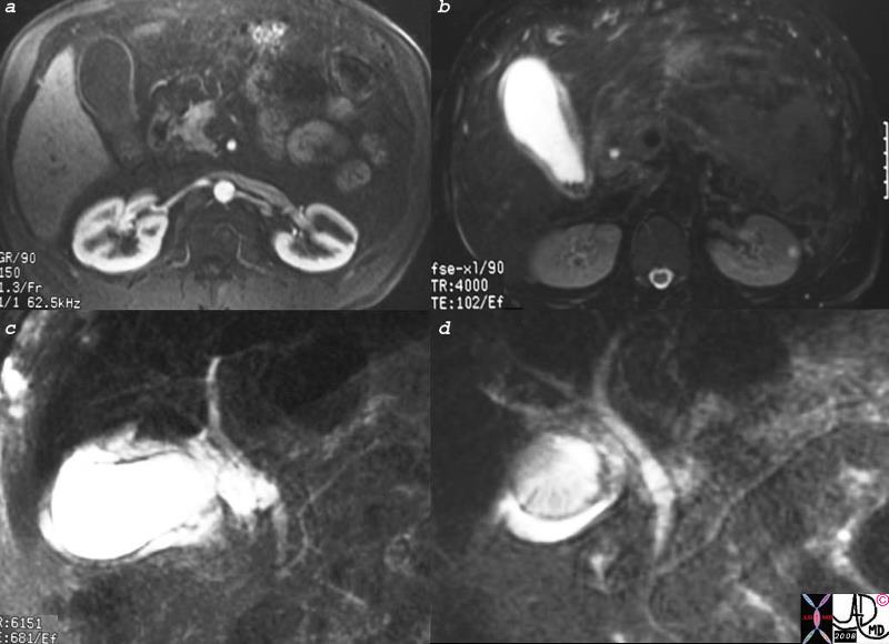
| Acute Renal Failure |
| 46787 hx 88 F with acute renal failure CT shoewed end stage left kidney and hydronephrosis on the right kidney renal pelvis ureter fx filling defect ureterolithiasis with proximal ureteral obstruction s/p nephrostomy radiologists and detectives – radiologists fingers pushing the catr=”theter” into better position elderly patient with compression fractures Davidoff MD |
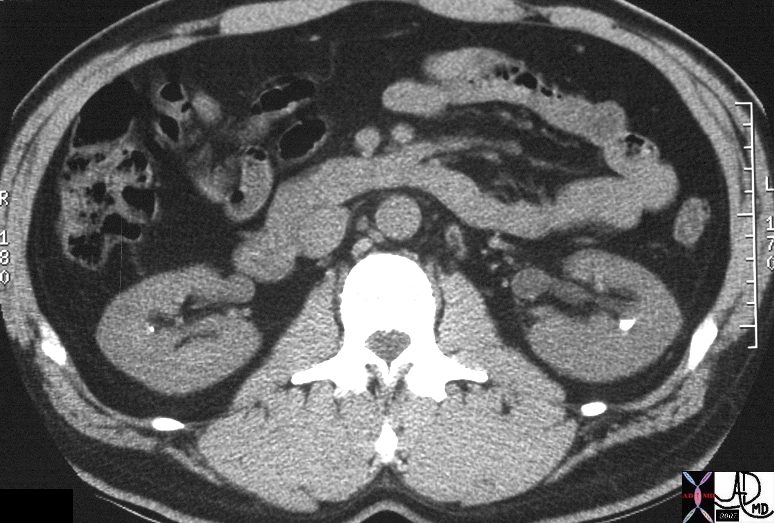
Nephrolithiasis and Milk of Calcium Urine |
| 16045 kidney renal nephrolithiasis fx calcifications calcified fluid fluid level dx milk of calcium urine CTscan Davidoff MD 16046b01 16046 |
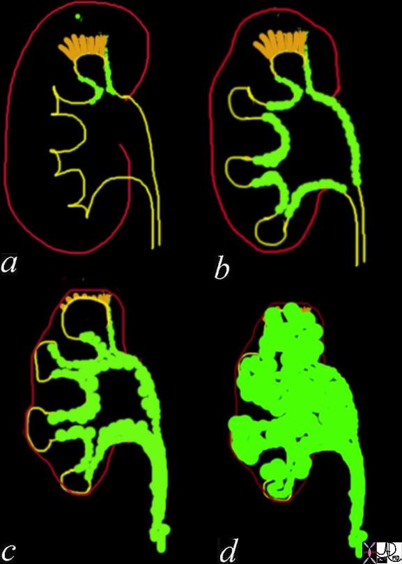
TB of the Kidney – Pathogenesis |
| 22419c01 kidney infection caseous necrosis infection TB tuberculosis autonephrectomy putty caseous necrosis calcification infundibular stenosis parenchymal infection submucosal spread sclerosis cicatrization hydronephrosis pressure atrophy Davidoff Art Davidoff Drawing Davidoff MD |
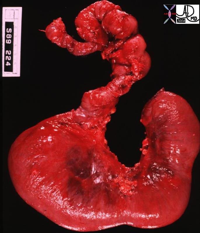
Dilated Small Bowel |
| 02062 small bowel fx dilated segment dx obstruction secondary to hypoplastic muscularis grosspathology |
Alternate Pathways in Disease – River Anaology |
| 01216 liver hepatic coronary vein esophageal varices dx portal hypertension cirrhosis transportal venogram venography Courtesy Ashley Davidoff MD |
Mechanical Disorders – Obstruction

Acute Ureteric Obstruction |
| 46610c01 kidney pelvic kdney renal position ureterolthiasis ureterovesical junction stone UVJ stone ureteric stone fx hydronephrosis fx etravasation dx acute obstruction of the right ureter from a 2mm calcified ureteric stone in a patient with a pelvic kidney CTscan Davidoff MD |
Occluded right bronchus |
| 46206 chest lung right mainstem bronchus fx occlusion dx aspiration CTscan Davidoff MD dx lobar atelectasis dx atelectatic RLL and LLL dx collapse due to bronchial obstruction |
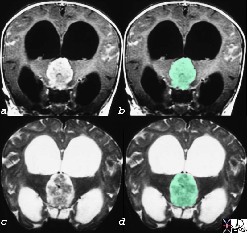
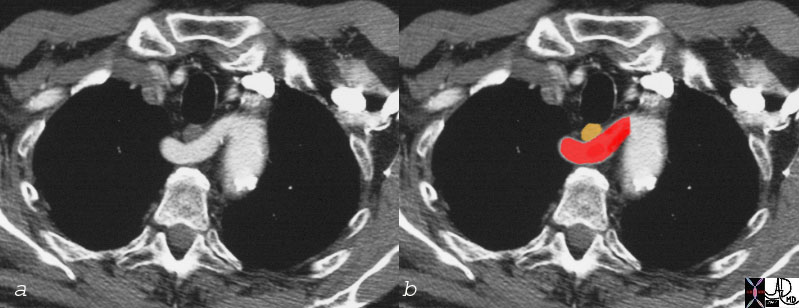
Papilloma of the Choroid Plexus Causing Hydrocephalus and Headache |
| The coronal MRI shows a gadolinium enhanced T1 weighted image (a,b) with black CSF in the dilated ventricles and a T2 weighted image with bright CSF (c,d). A large mass in the third ventricle (overlaid ingreen) dominates the study and is complicated by severe hydrocephalus. The mass was a choroid plexus papilloma of the third ventricle. In patients with brain tumor one of the causes of headache is hydrocephalus. This is an extreme case of hydrocephalus.
23223c01 head brain choroid plexus third ventricle hydrocephalus fx mass neoplasm papilloma MRI T1 T2 coronal with contrast C Courtesy James Donnelly MD |
Extrinsic Compression
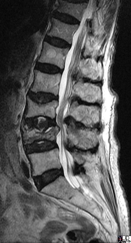 \ \
Compression of L4 with extrinsic effect on the Cauda Equina |
| 72021 bone vertebral body vertebra cauda equina lumbar spine CSF thecal space fx compression fracture extrinsic compression on thecal sac with displacement of the nerves of the cauda equina L4 |

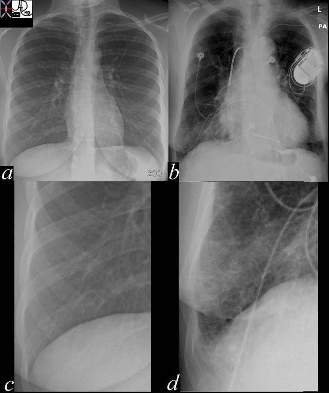
Left Upper Lobe Obstruction Collapse and Compensatory Forces |
| 49460c03 respiratory system lung left upper lobe fx atelecatsis compensation mediastinal shift lung shift volume loss plain CXR chest X-ray popcorn calcification Davidoff MD 49460c02.800 |
Incoordination
Penetration into the Vestibule |
| 76210c swallow penetration laryngeal vestibule hypopharynx barium swallow Courtesy Ashley Davidoff MD |

Aspiration |
| 46508c01.800 esophagus dysphagia fx large osteophyte causing difficulty with passing a scope fx aspiration barium swallow x-ray contrast Davidoff MD |
Mechanical Disorders – Degeneration

Wrath of Time |
| 46707c01 bone wrist carpals thumb degenerative changes osteoarthritis osteopenia age time normal vs abnormal X-ray plain film Davidoff MD |
Mechanical Disease – Loss of Elasticity
Normal Alveoli of the Lung and Emphysema
|
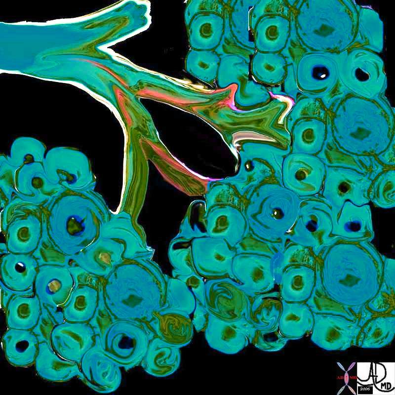
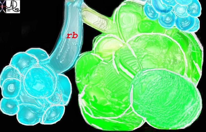
| This diagram illustrates the branching pattern of the tracheobronchial tree that extends from the bronchi to the terminal bronchioles transitioning into the alveoli via the alveolar sacs. Courtesy Ashley Davidoff MD 32645b04b04
This diagram shows alveoli and respiratory bronchioles that are too large due to loss of elasticity, so that air cannot be moved efficiently through them This is a diagram of emphysema causing hyperinflated lungs lung volumes 32645b01.800 Davidoff art |
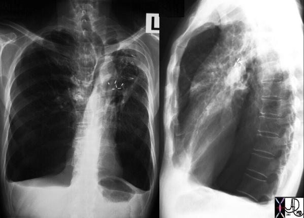
| Large lungs |
| A patient with hyperinflated lung volumes, COPD, and emphysema with surgical removal of a lung carcinoma from the LUL. Note how flattened the hemidiaphragms are and note the large retrosternal air space and the shapoe of the chest – like a barrel – called pectus carinatum – or pigeon chest. The lungs are literally so large that they are pushing the chest wall forward.
Courtesy Ashley Davidoff MD 30672c |
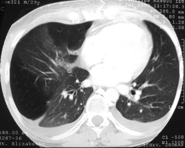
Segmental emphysema |
| This is a chest CT of a 29 year old man man who has segmental emphysema in the right lower lobe and midle lobe characterised by lucencies at 3 and 12 oclock where there has been air trapping in the alveoli. The left lung is normal This condition is called Swyer James syndrome Courtesy Ashley Davidoff MD. (30314 ) |
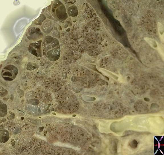
Emphysema |
| This is an image of an emphysematous lung. Note the larger air spaces where the septae between the alveoli, alveolar sac, salveolar ducts and respiratory bronchioles have been broken down. 19932e |
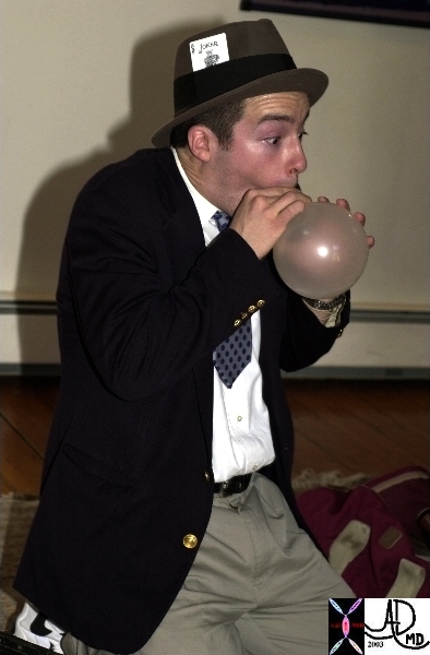
Pressure Tension Radius Laplace’s Law |
| This image shows a famous cardiologist, Dr Jonathan Fisher performing as Zippy the clown revealing and exaggerating the difficulty with surface tension and radius in the early stages of blowing up a balloon. Courtesy Ashley Davidoff MD. 60730b01 code lung physiology balloon surface tension surfactant alveolus alveoli radius principles |
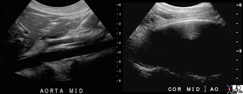
Normal aorta and Abdominal Aneurysm – Pressure Tension and Laplace’s Law |
| 70027c01 aorta abdomen abdominal aortic aneurysm normal aorta TCV applied biology AAA USscan Davidoff MD |
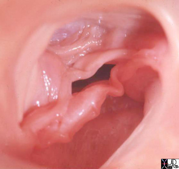
Thickened Bicuspid Aortic Valve |
| 07951b aorta aortic valve fx fusion of the commissures fx thickened bicuspid aortic valve congenital fused grosspathology Davidoff MD |
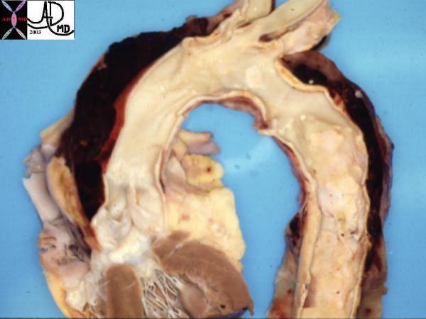
Dissection |
| This pathological specimen shows an aortic dissection starting at the root of the aorta and extending across the arch and into the descending portion. The false lumen is filled with clotted blood. Courtesy Henri Cuenoid MD 13421 code CVS thorax AO aorta dissection grosspathology |